What is it like to hold the beating heart of a two-day old child in your hand? What is it like to counsel distraught parents as they make some of the most difficult decisions of their lives?
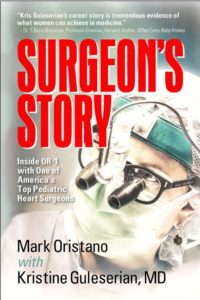
Noted pediatric heart surgeon Dr. Kristine Guleserian has opened up her OR, and her career, to author Mark Oristano to create Surgeon’s Story – Inside OR-6 With a top Pediatric Heart Surgeon.
Dr. Guleserian’s life, training and work are discussed in detail, framed around the incredibly dramatic story of a heart transplant operation for a two-year old girl whose own heart was rapidly dying. Author Mark Oristano takes readers inside the operating room to get a first-hand look at pediatric heart surgeries most doctors in America would never attempt.
That’s because Dr. Guleserian is recognized as one of the top pediatric heart surgeons in America, one of a very few who have performed a transplant on a one-week old baby. Dr. Guleserian (Goo-liss-AIR-ee-yan) provided her expertise, and Oristano furnished his writing skills, to produce A Surgeon’s Story.
As preparation to write this stirring book, Oristano spent hours inside the operating room at Children’s Medical Center in Dallas watching Guleserian perform actual surgeries that each day were life or death experiences. Readers will be with Dr. Guleserian on her rounds, meeting with parents, or in the Operating Room for a heart transplant.
Oristano is successful sportscaster and photographer and has made several appearances on stage as an actor. He wrote his first book A Sportscaster’s Guide to Watching Football: Decoding America’s Favorite Game, and continues to volunteer at Children’s Medical Center.
“We hear a lot about malpractice and failures in medical care,” says Oristanto, “but I want my readers to know that parts of the American health care system work brilliantly. And our health care system will work even better if more young women would enter science and medicine and experience the type of success Dr. Guleserian has attained.”
Readers will find all the drama, intensity, humor and compassion that they enjoy in their favorite fictionalized medical TV drama, but the actual accounts in Surgeon’s Story are even more compelling. One of the key characters in the book is 2-year-old Rylynn who was born with an often fatal disorder called Hypoplastic Left Heart Syndrome and was successfully treated by Dr. Guleserian.
A Day in the Life
“We eat stress like M&Ms in here.”
OR-5
Children’s Medical Center, Dallas
November 5, 2009
I’m staring at eleven month-old Claudia, lying sedated on the operating table in OR-5, as still as a doll with no moving parts. She looks smaller than her charted weight of nine kilos (20 pounds). Nurses cover her with sterile blue surgical drapes so all that’s visible is a 4-inch square patch of skin on her chest. Bright white lights bathe the center of the table. Doctors and nurses in gowns, caps, and masks crowd around. They look almost identical. Except for the earrings. The earrings are the “tell.” That’s how you know it’s her.
Kristine Guleserian, pediatric cardiothoracic surgeon, is scrubbed in. Known throughout the hospital as Dr. G, she is one of only nine women in the U.S. certified to do what she’s about to do — take a scalpel sharper than a dozen razors, cut through Claudia’s skin, saw open her breastbone, and spread her ribcage apart in order to repair two congenital defects threatening a malformed heart the size of a walnut. It’s just after 9:00 AM. Claudia will be in OR-5 until 2:00 PM, along with a team of talented surgeons, nurses, techs, anesthesiologists, and others. Dr. G is in charge.
October 27, 2009
Children’s Medical Center – Heart Center
Two weeks before Claudia’s surgery, I had a 1:30 PM meeting with Dr. G at her office. At 1:25, I sat in the waiting room. At 1:30, Dr. G came through at her favorite speed — full. She headed for the door while putting on her white, starched lab coat over surgical scrubs and said, “Come on.” We trotted down the hospital hallway.
“This is my world. You wanted to see it. Welcome to my life.”
“Where are we going?” I was struggling to keep up with her even though I’m a foot taller.
“We have to do a consult.”
“We?”
“I have to. You’ll watch.”
We whisked past the main desk of the echocardiography lab. Dr. G motioned to the charge nurse.
“He’s with me.”
We squeeze into the dark and cramped echo lab, where there’s barely enough space for the two women sitting at the monitors. Dr. G introduced me to cardiologists Dr. Catherine Ikemba and Dr. Reenu Eapen, then turned her focus to the echo monitors. An echocardiogram is a moving image produced by sound waves directed at the heart and reflected back again as the waves pass from one type of tissue to another. To me it looked like a blurry, moving x-ray. To the eyes of these three women it was an intimate cardiac road map. A nine-year old boy had a malformed aorta, and the cardiologists wanted Dr. G’s opinion. She was Socratic, asking questions she likely already knew the answers to, saying, “Well, I might do…” so-and-so, and then asking her colleagues for their opinions.
Two weeks later, I came back for the first of many long days as her shadow. I wasn’t quite Alice in Wonderland, but the feeling of falling down a hole did occur to me.
November 5, 2009
7:30 AM – Heart Center Research Meeting
There’s more to being a surgeon than surgery. This particular day begins in a windowless media room, the kind of video-meeting-training center you’d find in any school or business. Rows of desks and chairs give it a classroom feel. A/V equipment hangs from the ceiling and a large video screen dominates the front of the room. The dress code is strictly medical, no business attire here. Doctors and nurses in scrubs and lab coats shuffle into the room, many with the ubiquitous cup of Starbucks in hand. Today will feature a presentation of two ongoing cardiac studies being conducted at the Children’s Medical Center’s Heart Center. The room is very cold, and Dr. G wears a black turtleneck sweater under her white lab coat. She pulls the sweater neck up over her nose and mouth as the meeting goes on, seeking warmth. A presenter advances to the lectern, and the unmistakable look of the PowerPoint presentation flashes on the screen behind her. The title slide reads:
CHROMOSOMAL COPY NUMBERS IN
HYPOPLASTIC LEFT HEART SYNDROME
Before I ventured into Dr. G’s world, I had begun my own rudimentary study of congenital heart disease (heart defects present at birth), trying for a foothold in the maze of childhood cardiac problems. I had read that hypoplastic left heart syndrome (HLHS) is a life-threatening cardiac deformity where the left ventricle, which pumps blood to the aorta and then around the body, is so weak that without surgical intervention any infant suffering from it will likely die. The pediatric heart specialists in the meeting room critique what they’ve just heard. A senior cardiologist might question the validity of this or that portion of the research methodology. These are works in progress, not ready for publication. Ongoing study is a part of the surgeon’s job description.
In the meeting room, the media screen glows again.
ECHOCARDIOGRAPHIC PREDICTION OF SPONTANEOUS
CLOSURE OF DUCTUS ARTERIOSUS IN PREMATURE INFANTS
After only two weeks shadowing Dr. G, I was able to make some sense of this title. The Heart Center team is using echocardiography to predict whether the ductus arteriosus in the hearts of premature infants will close properly after birth, sparing the need for open-heart surgery. That was about all I knew. I had to dig deeper into the textbooks to learn more about what was beating beneath my own breastbone.
The human heart is a four-chambered pump, designed to send deoxygenated blood to the lungs to get a new supply of oxygen, and then sending that oxygen rich blood on its journey around the body to nourish organs and tissues. The left and right sides of the heart each have two chambers — an atrium on top, and a slightly larger ventricle on the bottom. Each side is like Dali’s version of an hourglass. The atria and the ventricles are each separated by a thin wall called a septum. The ventricular septum is slightly more muscular than the septum for the smaller atria.
In a normal heart deoxygenated (blue) blood enters the right atrium from large blood vessels called the vena cavae, which bring blood back from the rest of the body after distributing oxygen. The right atrium contracts, opening the tricuspid valve, and blood flows down into the larger right ventricle. The contraction of the right ventricle sends blood through the pulmonary valve to the pulmonary arteries, and into the lungs for oxygenation. The newly oxygenated blood enters the left atrium through the pulmonary veins. When the left atrium contracts, blood is sent through the mitral valve into the left ventricle. The left ventricle contracts, blood moves through the aortic valve into the aorta, and off to oxygenate the rest of the body — the brain, the coronary arteries of the heart itself, deep into the internal organs, and superficially to the skin. Over and over again, on average 100,000 times per day. That’s in an anatomically correct heart. (Anatomic trivia: The pulmonary arteries are the only arteries that handle deoxygenated blood, while the pulmonary veins are the only veins that handle oxygenated blood. Otherwise, oxygenated blood always flows through arteries, and deoxygenated blood through veins.)
The number of things that can go wrong with the human heart is staggering. Heart disease in adults is usually acquired. When we develop a heart condition in later life, it’s most often our own doing. Smoking, obesity, hypertension, poor diet, lack of exercise, diabetes, genetics and more, contribute to the clogged coronary arteries, heart attacks, strokes and other events that make heart disease the leading cause of death in most developed countries. Congenital heart disease is present in approximately 35,000 newborns in the U.S. each year, although many of these show no symptoms and don’t learn of any problems until years later, if ever. Since infants haven’t had a chance to do much damage to themselves, it’s fair to wonder how a newborn heart can have so many problems. Congenital heart defects occur because of interruptions in normal fetal heart development.
The developing fetal heart contains a series of shunts, like miniature bypasses, to keep blood away from the pulmonary arteries and lungs so that blood flow is kept low, and the tiny lungs won’t be overtaxed. Fetal lungs are non-functional, because the fetus gets oxygen from the mother through the umbilical cord. The shunts in the fetal heart are:
1) foramen ovale, which lets blood flow from the right to the left atrium,
2) ductus venosus, which draws umbilical blood away from the fetal lungs and into the vena cava, and;
3) ductus arteriosus, which connects the pulmonary artery to the descending aorta, thus allowing most blood from the right ventricle to bypass the non-functional fetal lungs.
All three of the shunts alter themselves after birth to create the normal heart design. When something interferes with the natural switch over from fetal to breathing infant heart, physicians call it “persistent fetal circulation.” It can manifest in hundreds of way. In certain situations, it’s never even noticed.
Anatomy of the Heart 101 is over. Bookmark these diagrams and return PRN (medical for “as needed”).
8:15 AM
3rd floor Cardiovascular Intensive Care Unit
The Cardiovascular Intensive Care Unit (CVICU) has twenty rooms arcing around a large central desk. The furnishings are modern, corporate-like, and austere. The pulse of the CVICU is the rhythm of the beeping sound common to every TV medical drama. Each patient is attached to a monitor measuring blood-oxygen saturation (sats), heart rate (HR), blood pressure (BP), respiratory rate, temperature, and more. Each monitor is a computer, producing different sounds for different reasons. The one constant is that audible beep, one for each heartbeat. An infant’s tiny heart beats significantly faster than an adult’s, so the pace of the beeping is rapid, and each baby here suffers from a potentially fatal malfunction of that rapidly beating heart.
Nurses move everywhere, monitoring every child. Intravenous (IV) fluid bags hang at each bed — six, eight, sometimes more. One patient has ten IV drips, each one delivering a different life-supporting medication — sedation, painkillers, antibiotics, anticoagulants, blood products, nutrition and others. The drips hang from poles, and flow directly into the tiny patient’s arm or leg, or more often, into a catheter inserted into the chest for easy access. The drips feed into a large control panel with the concentration and rate of flow of each drip handled by computer. All these babies are critically ill, critically tiny, many premature. Most of them are smaller than the stuffed animals that sit, unnoticed, next to them.
I’ve been volunteering at Children’s for 13 years, but this is my first time in the CVICU. I’m here for cardiac surgery rounds, following Dr. G as she checks on the progress of patients. Another familiar sight from medical TV shows is on display here — the long, white coat — the peacock feathers of physicians and surgeons. Children’s Medical Center is a teaching hospital, part of the University of Texas Southwestern Medical School in Dallas. Doctors and surgeons, long past their residencies now and specialists in their fields, wear the long, white lab coat. Medical students, residents and interns are in shorter coats. Dr. G is the shortest of the long coat-clad. Sure, she’s only five feet tall, but as they say in the sports world, she plays six-two. She’s not the only woman in the group, but she’s the only one wearing a long white coat. The young doctors listen to her.
Heart surgeons, ICU doctors, cardiologists, nurses, nurse practitioners, physician assistants, fellows, residents and students start at one end of the unit to move room by room around the floor. A cardiology fellow pushes the computer on wheels (COW), and presents each case. This young doctor has made several of the basic choices his career path requires. He’s just finished his residency where he worked in various specialties. He’s chosen medicine over surgery, pediatrics over adult, and cardiology over other disciplines, making pediatric cardiology his career choice. He’s taking his first steps down the six-year road it will take to earn “attending” status, when he’ll be in charge of cases. He’ll then be a pediatric cardiologist, a doctor who treats young people with heart disease. He’ll refer cases needing surgery to people like Dr. G, a pediatric cardiothoracic surgeon. Her career path was twice as long, requiring twelve years to attending status. Cardiologists diagnose — surgeons repair.
Even though he’s out of residency, this doctor is still learning. He stops in front of the door to the first patient room and runs down the important events from overnight — vital signs, patient status, complications, and planned treatment. The male attendings ask questions that are pointed and occasionally harsh. Dr. G draws the younger doctors out with her questions, gently nudging them back on the right track. “I didn’t hear anything about left atrial pressure there,” she tells the presenter, who immediately refers to the COW screen and spews a series of numbers out in a specific order. The young doctor’s voice is tense, rising a bit, as he makes up for his omission. It’s unlikely he’ll make this mistake again. Terms like “open-chest” and “life-threatening event” are heard on cardiac rounds, said calmly and with nonchalance. Hospital personnel in critical care settings are outwardly detached. It’s a key to staying focused.
The CVICU nurses rounding make notes while answering questions concerning how patients fared overnight. There is a pecking order among hospital personnel, and some doctors treat nurses as underlings; nevertheless, a tremendous level of trust exists between the doctors and nurses at Children’s. If the doctors are the officers of this army, the nurses are the sergeants, the ones who make sure everything gets done.
While the rest of the group moves along the hallway, Dr. G stops to look inside the room of the patient just presented. If she sees a family member inside, and they’re awake at this early hour, she goes in to say hello and ask how things are going. She feels a responsibility toward every family, even if the case isn’t hers. It’s not done for effect or because her medical training requires it. This is the way she treats everybody. It doesn’t matter if your child has a serious heart condition. It doesn’t even matter if you have a child. When Dr. G sees you, in the hallway, in the cafeteria, in the OR, she says hello.
Rounds end, leaving just enough time to dash up to the eighth floor cardiac unit and check on patients who are out of ICU, waiting to be discharged. One young heart transplant patient has turned up her oxygen level without the nurses knowing about it. Dr. G tells the 13-year old girl, in a firm, motherly way, that medical decisions are made by the pros and here’s how we’ll manage the oxygen for the remainder of your stay. The girl hangs her head and nods.
The moments after rounds, before the next issue presents itself, offer a chance to head down to the first floor food court for a snack. As Dr. G stands in the register line, her pager beeps. She checks the number and heads up to the third floor office suite she shares with her partners and staff. She phones the person who paged her and, in a flash, it’s out the door and back to the echo lab, a half-eaten banana left behind on her desk.
Two weeks after my first visit to the echo lab I stood to the side again, this time better able to make sense of some of what Dr. G and the cardiologists discussed as they looked at the screen. Eleven-month old Claudia’s diagnosis was Tetralogy of Fallot (TOF), a syndrome with four separate cardiac abnormalities:
1) Ventricular septal defect (VSD) — a hole in the wall between the two ventricles;
2) Overriding aorta — the aorta is not positioned properly on the heart;
3) Right ventricular outflow tract obstruction — for any of several possible reasons, the blood flow to the lungs is restricted, leading to:
4) Right ventricular hypertrophy, (which surgeons pronounce “hy-PER-tro-phy”) — a dangerous buildup of the right ventricle’s musculature.
Claudia has alarming episodes of cyanosis where her lips, fingers and toes turn blue because her oxygen saturation rate becomes dangerously low. She also has what are called “Tet spells,” when her oxygen level drops so low that she loses consciousness. The preoperative indications of most concern to Dr. G are an extremely small pulmonary valve, which leads from the right ventricle to the pulmonary arteries; the significantly thickened muscle bundle below the valve; and the somewhat larger than average VSD.
Thirty minutes later we were walking down a second floor hallway toward the operating rooms. Dr. G walked quickly, straight ahead, focused. She was getting her game-face on.
10:30 AM
OR 5
Claudia lay motionless on the table in the center of the OR, her head sticking through a hole in the draping around her neck. It’s visible to the anesthesiologists seated at the head of the table where they are concerned with the numerous gauges, medicines, inhalation gases and monitors at their fingertips. They’re also in charge of tilting the table at the surgeon’s request, to put the patient at a more favorable angle, because the motorized table can be raised, lowered and tilted to various angles at the touch of a button.
(Example of pediatric cardiothoracic humor —A flight attendant goes on the p.a. and asks if there’s a pediatric cardiac anesthesiologist on the plane. There is one, in the rear of coach. He signals the attendant and asks what the trouble is.
“There’s a pediatric heart surgeon in first class. He wants his tray table lowered.”)
The scrub tech stands at the opposite end of the table, facing a series of trays that hold an array of odd looking tools; forceps for picking up or grasping things; scalpels that slice through human flesh as if it were air; sutures (thread) finer than human hair, attached to small needles curved like fish hooks. The scrub tech is the right hand person to the surgeon, responsible for pulling instruments and supplies for the operation, knowing what the order of the operation is, and arranging everything in the most efficient format for this particular surgery and this particular surgeon. Dr. G knows that when she calls for an instrument, the proper one will be there in a flash. Often, it will be offered to her before she has to make the call.
A six-foot-by-six-foot metal frame sits to one side of the operating table, containing gauges, canisters, and clear plastic hoses. This is the cardiopulmonary bypass machine —“the Pump.” This technology will serve as Claudia’s circulatory system while her heart is stopped for repairs. Developed in the 1950’s, modern bypass machines still use hoses much like the beer keg tubing in the first experimental models. The two specialists in charge of operating the pump, the perfusionists, sit at the machine.
The small patch of Claudia’s chest that’s visible is covered with a material called Ioban, plastic coated with iodine in a further effort to reduce any risk of infection during surgery. Dr. G will make a tiny incision to get at this heart that was compromised in utero by Tetralogy of Fallot. To give you an idea of the progress of medical knowledge, TOF was first medically described, though primitively, in 1672. Two hundred years later Etienne Louis Fallot, a French physician, described the clinical pathology of the condition, but the first surgical treatment for TOF wasn’t available until the late 1940’s. Dr. G, ever the teacher, drew a diagram of the surgery for me before she scrubbed in.
After scrubbing, Dr. G re-enters the OR with hands and forearms still wet. She dries with sterile towels provided by a scrub tech who then helps her into a surgical gown and gloves. She wears loupes over her cap. They look like small telescopes growing from each eye, and they give her a magnified view of the tiny area in which she’s working. A fiber-optic cable runs up her back, over the top of her cap and onto a small, bright lighting instrument/video camera at her forehead, to light and televise what she sees to monitors hung around the OR. Dr. G is at the center of the sterile area, where only those who scrub in can go. The rest of us, wearing surgical masks and caps in addition to our scrubs, have to stay away from the table. She climbs up on a small step stool to get her five-foot frame high enough above the table to work easily, without making her taller assistants bend over.
She takes a scalpel and makes a four cm incision in Claudia’s chest. Next, she cuts the breastbone open with a small saw and puts retractors in place to hold the ribs apart. The first object Dr. G encounters inside Claudia’s chest is the thymus gland, a small, flesh-colored organ. It has some minor involvement with the lymphatic system, but it gets in the way of open-heart surgery, and you can live without it. So the gland is removed and discarded.
Dr. G takes an electronic scalpel called a “Bovie,” which cauterizes as it moves through tissue, keeping bleeding to a minimum. She cuts the pericardium, the sac-like membrane containing fluid that lubricates the heart. The pericardium has extra meaning for Claudia. Dr. G precisely excises a small portion of the sac and places it in a dish containing 0.6% glutaraldehyde, a preservative fluid. She’ll use this patch later to close the VSD, the hole between Claudia’s ventricles that failed to seal itself properly at birth. She works around the small space filled with tiny body parts, freeing up the aorta and the pulmonary arteries from the underlying tissue. Claudia has been given heparin, an anticoagulant, so that her blood is less likely to clot when it goes through the pump. Dr. G inserts cannulae, small tubes, into the aorta and the vena cavae. The other ends of these tubes are attached to the pump, connecting to Claudia’s circulatory system. Because Claudia has very small blood vessels, the work is delicate and precise, and the tubes they need for this bypass, like the vessels in Claudia’s chest, are extremely narrow. Her cannulae are smaller than the width of a ballpoint pen.
The mood in the OR shifts at various moments. Dr. G has been casually introducing me to the OR team while routine work is going on — as routine as heart surgery can be. But when the cutting starts, the room goes quiet. Dr. G hovers over the small body on the table, staring down into the chest she has cut open. The view from the camera attached to her loupes doesn’t shake on the OR monitors. She’s a human tripod.
The perfusionists are cooling Claudia’s body down to 28 degrees Celsius, 82.4 Fahrenheit, to slow her metabolism and protect her heart. Hypothermia lowers the amount of oxygen the brain requires, giving the surgeons time to perform the needed repairs. They aid this chilling process by turning the temperature in the OR down to 64 degrees, so cold that several people drape their shoulders with blankets from a nearby warmer.
Dr. G clamps the aorta, and blood stops flowing to Claudia’s heart. Dr. G tells the perfusionists to run the cardioplegia, a solution of chemicals inducing cardiac arrest. In order to operate on the heart they must intentionally cause something that usually kills when it happens on its own. The cardioplegia solution includes potassium chloride, one of the chemicals used in lethal injection executions. Claudia’s heart stops beating and the blood exits her vena cavae into the bypass machine for oxygen, returning to her body through the cannula inserted just above the clamp on the aorta. Her heart and lungs have been turned off. There’s no more beeping or EKG activity on her monitor. She has flat-lined. When the patient goes on pump the heart is like a water balloon with the water let out. It changes in shape from full and throbbing to flat and motionless. The only way to repair Claudia’s heart is to stop it and empty it.
The first task is to examine the heart to see if the preoperative diagnosis is correct. Dr. G uses delicate instruments to retract portions of the tricuspid valve and examine the extent of the defect of the ventricular septum, the wall between the two ventricles. She determines the exact size and shape of the VSD and trims the segment of pericardium she saved earlier in preservative. She cuts miniscule pieces of the pericardial tissue and sutures them along the walls of the VSD, creating anchor points for the actual covering. Each suturing is an intricate dance of fingers and forceps, needle and thread. Dr. G works with a small, hooked needle, grasping it with forceps, inserting the needle through the tissue, releasing and re-gripping with the forceps, pulling the hair-thin suture through, using a forceps in her other hand to re-grip the needle again and repeat. The pericardial tissue being sewn over the VSD has to be secure, and it has to stand up to the pressure of blood pumping through Claudia’s heart at the end of the operation. This isn’t like repairing knee ligaments, which can rest without use and heal slowly. Claudia’s heart is going to restart at the end of this operation, and whatever has been sewn into it has to hold, and work, the first time. The VSD repair involves cautious work around the tricuspid valve, and their proximity is a concern because the valve opens and closes along the ventricular septum with each beat. Dr. G and her team find that it’s preferable to actually divide the cords of the tricuspid valve to better expose the VSD. After the patch is fully secured, the tricuspid valve is repaired.
Things don’t go as smoothly during the attempt to repair the pulmonary valve. When Dr. G looks inside Claudia’s heart she discovers that the pulmonary valve is not nearly large enough, and it’s malformed. It only has two flaps where there should be three. She repairs it by what she later says is “just putting in a little transannular patch.”
Here’s what it’s like to “just” put a transannular patch on the pulmonary artery of a child as small as Claudia:
First, take a piece of well-cooked elbow macaroni. Tuck it away in a bowl of pasta that has a bit of residual marinara sauce still floating around in it. Take several different sized knitting needles. Slowly, without damaging the macaroni, insert one of the knitting needles into it to see if you can gauge the width of the macaroni on which you’re operating. Then using a delicate, incredibly sharp blade, cut a small hole in the piece of elbow macaroni, maybe a little larger than the height of one of the letters on the page in front of you. Now use pliers to pick up a small needle with thread as fine as human hair in it. Use another pliers to pick up a tiny piece of skin that looks like it was cut from an olive, so thin that light shines through it. Take the needle and sew the olive skin on to the hole you’ve cut in the piece of macaroni. When you’re finished sewing, hook up the piece of macaroni to a comparable size tube coming from the faucet on the kitchen sink, and see if you can run some water through the macaroni without the patch leaking.
That’s the food analogy. Those are the dimensions Dr. G worked with as she patched Claudia’s pulmonary artery. She made it a little wider to give it a chance to work more efficiently, to transport more blood with less blockage, requiring less work for the right ventricle so that the built-up heart muscle could return to a more normal size. It wasn’t the repair she’d planned to make, but it was the most suitable under the circumstances, and it gave Claudia her best chance.
Before restoring Claudia’s natural circulation, the team makes certain that no air is in the heart or the tubes from the pump, because it could be pumped up to the brain. Air in the brain is not a safe thing. When all the repairs are completed, Claudia is rewarmed and weaned from the bypass machine. She was on pump for 114 minutes and her aorta was clamped for 77 minutes, not an extraordinary length of time in either case.
Claudia’s heart starts up on its own, with a strong rhythm. With her heart beating again the beeps, and the peaks and valleys on her monitor return. All is well. An echo technician wheels a portable machine into the OR and puts a sensor down Claudia’s throat where it lodges behind her heart to perform a transesophageal echo —a more detailed view than the normal, external echo. Everything looks good. Chest drains are put in to handle post-operative drainage, and wires are placed for external pacemakers, should anything go wrong with Claudia’s heart rhythm during her recovery from surgery. Dr. G draws Claudia’s ribcage back together with stainless steel wires, perfectly fastened and tightly tucked down.
Claudia and the surgical team return to the CVICU, and Dr. G monitors her reentry to the unit, making sure the nurses understand Claudia’s condition and the proper procedures to be followed for the next 24 hours. From there, Dr. G enters a small room tucked away from the noise of the unit to meet with the family. Claudia’s mother, father, and aunt are waiting. Dr. G sees Mom wiping tears away.
“Are you crying? Oh, no, no need to be crying, everything is fine.” Her wide smile reassured Mom, who put away her tissues.
She tells the family what she did, and why she did it, using a serviceable mixture of medical and lay terms.
“I got in and saw the valve and it was really abnormal,” Dr. G tells the family, “really, really small. It only had two leaflets, and that’s not good, it’s supposed to have three. So I did a little transannular patch through a mini-sternotomy, which is really good for her — much smaller scar. Her echocardiogram was beautiful. There’s no hole where we closed her VSD. We know there’s another small, little hole in the muscle, but we all agreed that because it’s in the muscle it’s going to close on its own, so we won’t worry about it. My plan is, once she wakes up later today, to get the breathing tube out.”
There is a noticeable sense of relief evident on the family faces, even though one or two of the terms may have been unfamiliar. Then, comes the caveat.
“The arteries that go to each lung are a little bit small. She’ll need to have a pulmonary valve at some point. Some people need one not so long from now. Some people go a good portion of their lifetime without needing one. My brother had this same surgery when he was little, and he still hasn’t had a new valve put in yet. But he will some day.”
The simple fact that her brother had similar surgery seems to put the family a little more at ease. They know Dr. G has been on both sides of the equation, and she can relate to their anxiety.
From there it was off to a brainstorming session with the architects designing new cardiac surgery suites. They wanted staff input on what should go where, how far the doors should be from the operating tables, etc. In the OR, a matter of a few feet can mean the difference between life and death.
Lunch came at 3:30, which can actually be early in Dr. G’s world. She debriefed herself from the surgery as we ate, describing to me what had taken place. She would later dictate all this for the official surgery report in medical terms such as, “The right-sided pericardium was fenestrated to approximately 1 cm anterior to the right phrenic nerve.” It may be true that “the heart has reasons which reason knows not of.” It also has a language that’s pretty hard to understand as well.
I told Dr. G this was my first time in the OR and I couldn’t believe I’d just seen a kid’s heart beating inside her chest.
“You’ve never seen that before?” she asked me.
I reminded her that I’d spent the last 30 years as a sportscaster.
“It’s not exactly the kind of thing you see in the Dallas Cowboys locker room.”
She was genuinely surprised at my sense of wonder.
The rest of her day consisted of phone calls, emails, consults with other surgeons, afternoon rounds through the CVICU (which move more quickly than morning rounds, as these are just for checking up on each patient one more time), and the never-ending battle with paperwork.
On rounds at 7:30 tomorrow morning, Dr. G will check up on Claudia to see how she’s doing. That’s assuming she makes it through the night easily. If problems develop, it’s likely Dr. G could spend the night here with her.
“We eat stress like M&Ms in here,” said Dave Bartoo, her surgical tech this day.
This is where Dr. Kristine Guleserian repairs the tiny hearts of tiny children.
Come on in.
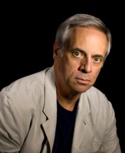
Mark Oristano has been a professional writer/journalist since the age of 16.
After growing up in suburban New York, Oristano moved to Texas in 1970 to attend Texas Christian University. A major in Mass Communications, Mark was hired by WFAA-TV in 1973 as a sports reporter, the start of a 30-year career covering the NFL and professional sports.
Mark has worked with notable broadcasters including Verne Lundquist, Oprah Winfrey and as a sportscaster for the Dallas Cowboys Radio Network and Houston Oilers Radio Network. He has covered Super Bowls and other major sports events throughout his career. He was part of Ron Chapman’s legendary morning show on KVIL-FM in Dallas for nearly 20 years.
In 2002 Oristano left broadcasting to pursue his creative interests, starting a portrait photography business and becoming involved in theater including summer productions with Shakespeare Dallas. He follows his daughter Stacey’s film career who has appeared in such shows as Friday Night Lights and Bunheads.
A veteran stage actor in Dallas, Mark Oristano was writer and performer for the acclaimed one-man show “And Crown Thy Good: A True Story of 9/11.”
In 1997, Mark began volunteering at Children’s Medical Center in Dallas, working in the day surgery recovery room. It was at Children’s that Mark got to know Kristine Guleserian, MD, first to discuss baseball, and later, to learn about the physiology, biology, and mystery of the human heart. That friendship led to a joint book project, Surgeon’s Story, about Kristine’s life and career.
Mark is married and has two adult children and two grandchildren.